
PUJ obstruction, also known as Pelvi-Ureteric Junction obstruction, is a condition where there is a blockage at the junction between the kidney (renal pelvis) and the ureter (the tube that carries urine to the bladder). This blockage prevents normal urine flow, leading to swelling of the kidney, a condition called hydronephrosis.
PUJ obstruction can be present at birth (congenital) or may develop later in childhood. Early diagnosis and timely management are important to protect kidney function.
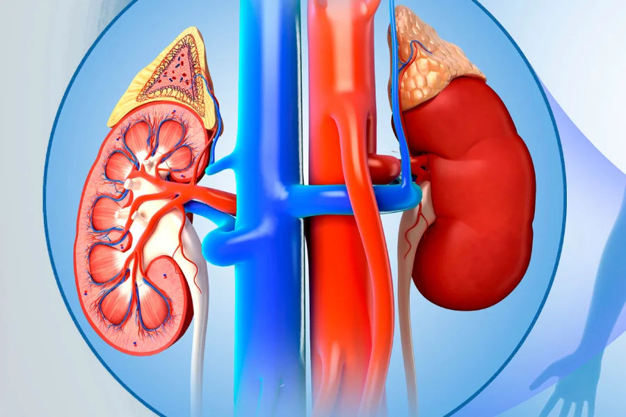
PUJ obstruction may occur due to:
Narrowing at the junction between the kidney and ureter
Abnormal muscle development at the junction
Crossing blood vessels compressing the ureter
Scar tissue (rarely in older children)
Most cases in children are congenital and detected during prenatal ultrasound.
In many infants, PUJ obstruction is detected before birth through routine pregnancy scans. In older children, symptoms may include:
Abdominal or flank pain
Recurrent urinary tract infections (UTIs)
Vomiting
Blood in urine (rare)
Swelling or lump in the abdomen
Some children may remain symptom-free and are diagnosed during evaluation for hydronephrosis.
Evaluation may include:
Ultrasound scan to detect kidney swelling
Renal scan (DTPA or MAG3 scan) to assess kidney function and drainage
Urine and blood tests
Additional imaging if required
These tests help determine the severity of obstruction and the need for treatment.
Treatment depends on the severity of the blockage and kidney function.
If kidney function is normal and swelling is mild, regular monitoring with ultrasound scans may be recommended. Some mild cases improve over time.
If the obstruction is significant or kidney function is affected, surgery is advised.
Pyeloplasty is the standard surgical procedure and involves:
Removing the narrowed segment
Reconnecting the healthy ureter to the kidney
Restoring normal urine flow
The surgery may be performed using:
Open surgery
Laparoscopic (minimally invasive) technique
Minimally invasive surgery offers smaller incisions, less pain, and quicker recovery.
A complete evaluation is done to assess kidney function. Parents are informed about the procedure and recovery process.
The procedure is performed under general anesthesia in a specialized pediatric surgical setting.
A temporary stent or drainage tube may be placed to aid healing. Most children recover well and resume normal activities within a few weeks. Follow-up scans ensure proper kidney function.
Consult a pediatric surgeon if your child has:
Persistent flank or abdominal pain
Recurrent urinary infections
Swelling in the abdomen
Known hydronephrosis detected on scan
Early treatment helps preserve kidney function and prevents long-term complications.
Our pediatric surgical team provides comprehensive evaluation and advanced treatment for PUJ obstruction. With precise surgical techniques and careful follow-up, we focus on protecting kidney health and ensuring safe, long-term outcomes for your child.